Knee Joint Injuries
The knee is the largest joint of the body, and also one of the most vulnerable to injury. It is made up of numerous structures, including bone, ligament, cartilage, muscle, tendon, and meniscus, all of which are essential to its function as a whole. In the event of a knee joint injury, it is important to have a good idea which structures are involved, and the severity of the damage, in order to determine which treatment options are best.
Anatomy of a Knee Injury
Bones
Three major leg bones come together to form the knee joint; the femur (aka thighbone), tibia (shinbone) and the patella (kneecap). The fibula, the outer and smaller lower leg bone, is also connected to the knee joint by the lateral collateral ligament (LCL). In the event of an injury, bones of the knee joint can be fractured or dislocated.
Fractures
A fall from a height or high-impact collision can cause one or more bones that make up the knee to crack, break into two, or shatter. The angle of the break will vary based on the injury event, and it is likely the soft tissue structures of the knee will also be damaged.
Dislocation
High-speed dynamic movements, and high-impact falls/collisions can cause the femur and tibia to move completely out of alignment. Dislocation can also occur when there is pre-existing knee instability or misalignment, due weakness or injury to surrounding muscle or ligaments. Note: A fracture or dislocation is a serious medical condition. Seek medical attention immediately, if you have any of these symptoms:
- Severe pain and swelling
- Bruising
- Pain with or inability to bend knee or straighten leg
- Knee or surrounding area appears misshapen
- Bone protruding from skin
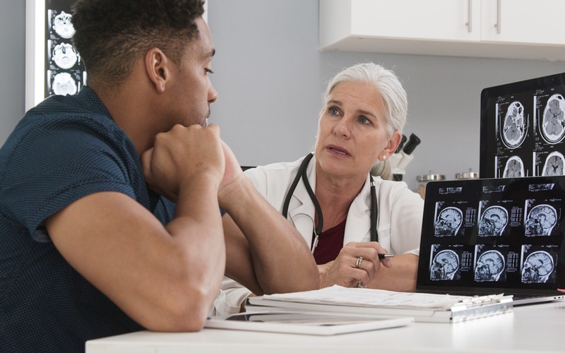
- X-ray and Computed Tomography (CT) scans are used to determine if there has been damage to bone tissue. These types of medical imaging use radiation to examine the condition and placement of internal body structures. Dense material, such as bone, is highlighted in the resulting image because it absorbs more of the x-ray radiation than less dense material in the surrounding area.
- Platelet-rich plasma (PRP) injections of the patient’s blood platelets may be used to reduce pain and can help speed up the healing process of soft tissues.
- Bracing, casting and splinting are non-surgical treatment options used by an orthopedic specialist to hold the bones in proper alignment, and to limit any extra movement, while they heal.
- Pain Management is an important component to any treatment and a long term rehabilitation program. In addition to PRP injections, other Orthopedic treatment options include cortisone shots which relieve pain and inflammation in target areas of the body.
- Physical therapy is necessary for restoring the strength, stability, and range of motion of the knee joint once the area is in the rehabilitation stage of recovery.
- External and Internal Fixation methods surgically insert pins, screws, wire, rods and/or metal plates into the affected and surrounding bone in order to facilitate correct alignment during the healing process. External fixation techniques, unlike internal fixation, utilize a metal framework of bars and screws which connect to those placed inside the bone.
- Total knee replacement or bone graft is needed if the bone is shattered into pieces too small to connect with pins or plates. During a bone graft procedure, bone tissue from another part of the body is transplanted to the damaged area. The cells from the replacement bone fuse to the damage bone. In a knee replacement, bone is replaced with artificial material.
Ligaments
The bones of the knee joint are held together by dense connective tissues known as ligaments. Ligaments keep joints stable by limiting the way in which bones move in relation to one another. The knee, as a modified hinge joint, is permitted to flex and extend (like a door hinge), and is also allowed to rotate inward and outward slightly. Ligaments are viscoelastic, which means they can lengthen to a certain extent when under stress, and then return back to their normal length when relaxed. However, if pushed beyond their normal range repeatedly or traumatically, ligaments can be permanently stretched or torn-also known as a sprain.
Anterior Cruciate Ligament (ACL) Sprain
The ACL connects the femur (thighbone) to the tibia (shinbone), and is a major knee joint stabilizer. It keeps the femur and tibia from sliding out of alignment during hinging and rotational motions. It sits under the patella (kneecap), and crosses the space between the femur and tibia at a diagonal. The ACL is one of four ligaments that form the knee joint, but is the most often damaged during sports-related activities. The severity of an ACL sprain is ranked from Grade 1 to Grade 3. Grade 3 describes a completely torn ACL.
- Sharp, severe pain in the knee
- Audible “popping” sound and sensation in the knee
- Immediate swelling
- Feeling unstable when putting weight on the knee
- Bruising
- Tenderness to the touch
- Loss of range of motion
- Mi-eye 2 Joint Arthroscopy is a hand held device equipped with a needle and a camera, and provides an in-office alternative to MRI, for those who are not MRI candidates. An orthopedic surgeon can view inside the joint cavity and make a diagnosis for treatment.
- Magnetic Resonance Imaging (MRI) uses powerful magnets to view soft tissue structures inside the body. If a ligament injury is suspected, a doctor will prescribe an MRI. However, an MRI is not always necessary to diagnose an ACL sprain.
- Platelet-rich plasma (PRP) injections reduce pain and can help speed up the healing process using the patient’s own blood platelets.
- Bracing is appropriate for low grade ACL injuries to patients with low activity lifestyles, such as older adults. More serious ACL sprains will require more intrusive treatment.
- Pain Management is an important component to any treatment and a long term rehabilitation program. In addition to PRP injections, other Orthopedic treatment options include cortisone shots which relieve pain and inflammation in target areas of the body.
- Physical therapy is necessary for restoring the strength, stability, and range of motion of the knee joint once the area is in the rehabilitation stage of recovery.
- Tissue grafts are used to help reconstruct the damaged ACL, and typically involve transplanting a portion of muscle tendon to bridge the gap, and help to direct the growth of scar tissue.
Meniscus and Tendons
Meniscus is a C-shaped piece of cartilage within the knee joint. The two menisci in each knee joint serve as the “shock absorbers” between the femur and tibia. Tendons connect muscle to bone. The tendons most directly associated with the knee are the patellar tendon and the quadriceps tendon. A tear to the meniscus or a tendon can occur with abrupt twisting, sudden changes in direction, high-impact falls, or from overuse. Degenerative disorders can cause meniscus to wear away over time.
- mi-eye 2 Joint Arthroscopy is a hand held device equipped with a needle and a camera, and provides an in-office alternative to MRI, for those who are not MRI candidates. An orthopedic surgeon can view inside the joint cavity and make a diagnosis for treatment.
- Magnetic Resonance Imaging (MRI) uses powerful magnets to view soft tissue structures inside the body. If a ligament injury is suspected, a doctor will prescribe an MRI.
- Rest, Ice, Compression, Elevation (RICE) procedure at home.
- Platelet-rich plasma (PRP) injections reduce pain and can help speed up the healing process using the patient’s own blood platelets.
- Bracing (immobilizing the knee)
- Pain Management is an important component to any treatment and a long term rehabilitation program. In addition to PRP injections, other Orthopedic treatment options include cortisone shots which relieve pain and inflammation in target areas of the body.
- Physical therapy is necessary for restoring the strength, stability, and range of motion of the knee joint once the area is in the rehabilitation stage of recovery.
- Tear repair involves suturing (sewing together) the ends of the meniscus, or the affected tendon edge to the top of patella (kneecap). With time, the tissue will regenerate.
- Meniscectomy is needed if the tear occurs in an area of the meniscus that does not receive blood supply (white meniscus). This is a portion of the meniscus that will not regenerate. In this procedure, the damaged tissue will be cut away. For appropriate cases, artificial cartilage is used to replace the meniscus.
Rotator Cuff Injuries
Anatomy of Rotator Cuff Injuries
Bones
Three bones meet at the shoulder joint: the humerus (upper arm bone), the scapula (shoulder blade) and the clavicle (collarbone). The shoulder joint is a type of synovial joint called a “ball and socket” joint, and is capable of the widest range of motion of any other joint type. The head of the humerus (upper arm bone) sits inside the glenoid fossa, which is a concave area of the scapula (shoulder blade). The shoulder is the most commonly dislocated joint, and many dislocations occur alongside bone fractures. The shoulder joint is especially susceptible to injury during high-contact sports such as hockey and football, but also during mountain biking and skiing. Damage to soft tissues such as tendons and cartilage will be present with fractures and joint dislocation.
Fractures
Fractures to the clavicle, humerus and scapula typically occur as the result of a direct blow to the area, as in a high-impact collision or fall from a height.
Disclocation
Dislocation of the shoulder describes the removal of the head of the humerus from the glenoid fossa. High-impact collisions can cause shoulder dislocation. The arm can also be pulled out-of-socket while climbing, and during high-speed rotation such as throwing a basketball
- Sharp, severe pain in the shoulder
- Immediate swelling and bruising
- Tenderness to the touch
- Loss of range of motion
X-ray and Computed Tomography (CT) scans are used to determine if there has been damage to bone tissue. These types of medical imaging use radiation to examine the condition and placement of internal body structures. Dense material, such as bone, is highlighted in the resulting image because it absorbs more of the x-ray radiation than less dense material in the surrounding area.
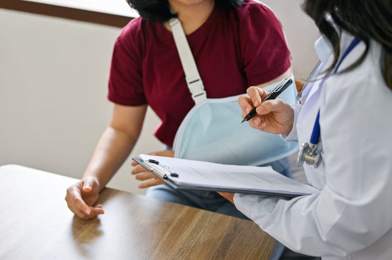
- Closed reduction or “setting” the broken bone is necessary for most shoulder fractures. If the skin is not broken, and if the break is mild, a healthcare practitioner will be able to put the bones back into the proper placement before immobilizing the joint with a cast, brace or sling.
- Platelet-rich plasma (PRP) injections of the patient’s blood platelets may be used to reduce pain and can help speed up the healing process of soft tissues.
- Bracing, casting and splinting are non-surgical treatment options used by an orthopedic specialist to hold the bones in proper alignment, and to limit any extra movement, while they heal.
- Pain Management is an important component to any treatment and a long term rehabilitation program. In addition to PRP injections, other Orthopedic treatment options include cortisone shots which relieve pain and inflammation in target areas of the body.
- Physical therapy is necessary for restoring the strength, stability, and range of motion of the shoulder joint once the area is in the rehabilitation stage of recovery.
- External and Internal Fixation methods surgically insert pins, screws, wire, rods and/or metal plates into the affected and surrounding bone in order to facilitate correct alignment during the healing process. External fixation techniques, unlike internal fixation, utilize a metal framework of bars and screws which connect to those placed inside the bone.
- Shoulder joint replacement may be necessary if the bones involved have been shattered and other surgical repair procedures are not appropriate.
Rotator Cuff Tendons
The muscles of the shoulder are referred to as the “rotator cuff muscles”. They are responsible for stabilizing the scapula and the head of the humerus in the glenoid fossa. Rotator cuff tears are seen in tennis and baseball due to overuse, but can also occur as a result of injury.
- Pain in the shoulder during movement, especially moving the arm above the head and behind the back.
- Stiffness and weakness of the arm and shoulder
- Inflammation around the joint
- Tenderness to the touch
- Loss of range of motion
- Popping sound or sensation with movement
- Physical assessment with range of motion test
- mi-eye 2 Joint Arthroscopy is a hand held device equipped with a needle and a camera, and provides an in-office alternative to MRI, for those who are not MRI candidates. An orthopedic surgeon can view inside the joint cavity and make a diagnosis for treatment.
- Magnetic Resonance Imaging (MRI) uses powerful magnets to view soft tissue structures inside the body.
- Rest, Ice, Compression, Elevation (RICE) procedure at home.
- Platelet-rich plasma (PRP) injections reduce pain and can help speed up the healing process using the patient’s own blood platelets.
- Sling (immobilizing the shoulder)
- Pain Management is an important component to any treatment and a long term rehabilitation program. In addition to PRP injections, other Orthopedic treatment options include cortisone shots which relieve pain and inflammation in target areas of the body.
- Physical therapy is necessary for restoring the strength, stability, and range of motion of the knee joint once the area is in the rehabilitation stage of recovery.
- Tear repair involves reattaching the tendon to the humerus with sutures. With time, the tissue will regenerate.
- Tendon transplant is necessary if there is not enough of the original tendon to attach to the bone. An Orthopedic surgeon will use a nearby portion of tendon as a replacement.
Carpal Tunnel Syndrome
Anatomy of Carpal Tunnel Syndrome
Carpal Tunnel Syndrome occurs when the median nerve, which is surrounded and protected by the carpal tunnel, in the wrist, is compressed by inflamed flexor tendons and ligaments. The top of the tunnel is made up of the transverse carpal ligament. The median nerve is responsible for transmitting electrical signals from the brain to the many parts of the hand. The syndrome is an infamous problem for people who engage in repetitive fine motor work activities or hobbies. Frequent pressure and stress to the wrist and palm area is also a common factor in gymnastics and yoga, as well as any other sports activities that involve grasping and extreme wrist flexion or extension. The wrist is also often vulnerable to soft tissue tears and fractures. Carpal Tunnel Syndrome can occur in a wrist healing from acute injury.
- Numbness and tingling in the fingers
- Pain in the thumb, index, middle and ring fingers
- Weakness in the hand, especially when trying to grab, pick up, or complete fine motor tasks
- Physical performance tests assess hand strength and responsiveness
- Electromyogram (EMG) and Nerve Conduction Velocity (NCV) tests are performed by Orthopedic specialists to measure the degree to which nerve impulses are able to communicate throughout the target area, and to the rest of the body.
- X-ray and Computed Tomography (CT) scans are used to determine if there has been damage to bone tissue. These types of medical imaging use radiation to examine the condition and placement of internal body structures. Dense material, such as bone, is highlighted in the resulting image because it absorbs more of the x-ray radiation than less dense material in the surrounding area.
- Magnetic Resonance Imaging (MRI) uses powerful magnets to view soft tissue structures inside the body.
- Splinting or Bracing (immobilizing the wrist)
- Platelet-rich plasma (PRP) injections reduce pain and can help speed up the healing process using the patient’s own blood platelets.
- In addition to PRP injections, other Orthopedic treatment options include cortisone shots which relieve pain and inflammation in target areas of the body.
- Physical therapy is necessary for restoring the strength, stability, and range of motion of the knee joint once the area is in the rehabilitation stage of recovery.


